User Tools

Learn about Electron Microscopes. What's an Electron Microscope? By Royce Barber. Please let me know if this all makes sense. REVISED Aug-5-2017 to add “Scanning VS Transmission”.
.
.
.
.
.
.
Anyone can understand! Also a bit about X-Rays and radiation. We'll first talk about the super popular “SEM” Scanning Electron Microscope. That just means it scans.
.
Simply said, the scanning electron microscope feels in the dark, then it visually shows you a accurate-enough representation of what it felt. Its feelers are very small. Without color. It shows you a drawing it makes of the object. Like if you feel a brick wall in the dark, you get a black and white idea of what the wall looks like. The microscope shoots tiny tennisballs into a dark box, and the way the ball bounces back, gives an idea of what the ball was hitting. Rapidly shooting a stream of tennis back and forth along a super-bumpy wall, and you'll have enough data to know what that wall looks like, because you'll only be seeing the balls that bounce back. The tennisballs have to be smaller than most cracks in the wall. So the wall has to be pretty bumpy to get a great image. Each tennisball is actually an electron, shot out of a gun and then aligned using magnets. Then the electron bounces off your prepared sample and that bounce-back is examined. Each bounce, basically shows up as one pixel on your computer screen.
.
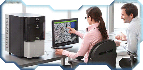
Before I explain the process in more detail, here's what you do with the microscope. An electron microscope comes in different shapes and sizes, all using a similar approach. They help in seeing what nearly anything is made of, at a far higher detail than the magnifying-glass microscopes you used in middleschool. Magnifying-glasses distort images and the light goes all over the place, making the image warped and blurry. The more lenses, the more the light is blurry and has hazy halo-like colors in it. Electron Microscopes (all major kinds) don't need light or magnifying glasses. You can experiment and create new kinds of metals, because the electron microscope lets you see how strong chemicals bond together, and what structures look like, so you'll know the close-up results of mixing metals. Electron Microscopes are vital to producing computer processors. Some newer electron microscopes are as small as a desktop computer, but still cost as much as a new car. The SEC “Scanning Electron Microscope” is highly popular and often easy to use. A small group of people can chip in to purchase a used electron microscope the size of a small car.
.
Now let's get started using any electron microscope. First you use a small shoe-box sized machine called a Sputter, to clean and spray a sample you wish to examine. The sample is coated with a super-thin metal which is highly visible to your electron microscope. The spray is needed, because the microscope is terrible at seeing without it. Some people place the sample in multiple Sputter machines to clean and coat in various ways, depending on the quality desired. The spray also protects the sample later on when the microscope is heating up.
.
Now you place your protected sample on a tray and into the Scanning Electron Microscope. Dusty room-air is pumped out, and super-frozen gas is added. A metal wire is heated red hot, and naturally shoots electrons. (Did you know, a metal coat-hanger can levitate if you apply a lot of electricity? It shoots out the electricity all over the place). The electrons shot out of the metal wire are messy, and so a magnet pulls them into a steady beam, and onto exact places on your sample. The electrons bounce off your sample, and then is attracted into a sensor which rapidly studies the direction/energy/speed/condition of each electron. Electrons are shot at your sample in different areas, in a grid, and it shows up on your screen as light or dark pixels. Making a topography map.
.
Remember, if you close your eyes and feel and object, you will get an idea of the shape and hardness of any object. The areas you felt, show up in your brain as a shape. The Electron Microscope feels an object by shooting electrons at it, and watching how the electrons bounce away from the shape.
.
You can generate a 3D model of your sample, by shooting electrons at it from different directions. Sadly it won't be in color, due to light reacting strange at such small sizes. Light looks weird up close. Groups of objects can be colorized by your computer, and sometimes groups of chemicals can be colorized. Sometimes a light-through-magnifying-glass can be used to get a very rough idea of the color of objects at a distance, and to give you a general map of the entire sample so you can navigate your electron microscope.
.
The sample can be rotated and tilted. You can even see two chemicals reacting on an atomic level, and set the microscope to take a image every few seconds or hours or days. You may make a time-lapse animation of chemicals interacting.
.
Many Electron Microscopes also have optional X-Ray features, to look inside the object, but it's often not a pretty image when many layers are blended together. How X-Ray works is another story involving radiation shooting through soft materials and somewhat through some dense materials. Radiation is electromagnetic waves, or very-very small objects that pass through a wall like water through mesh fabric. Side note, the TSA at airports, can 3D-scan your backpack using the same mild radiation your dentist uses to see through human tissue. If you hold a bright light to one side of your hand, you'll see the glow of red blood through your hand, because light penetrates soft tissue. Sun radiation can easily cook human tissue, so your hair and clothing keep you safe.
.
.
.

.
.
.
Another kind of Electron Microscope? We went over how the super-popular “SEM” Scanning Electron Microscope bounces electrons off your sample. Now we'll mention the other kind of Electron Microscope.
.
The “TEM” Transmission Electron Microscope. Not as popular but higher resolution. Instead of Electrons bouncing off the protected sample, the electrons shoot through the sample (like water through cloth), which often damages the sample badly. Electrons burn and bruise the sample as they travel through open gaps. Samples sensitive to radiation are quickly torn and damaged! Untreated biological material (your skin) is extremely sensitive to radiation, has low contrast (difficult to see), and doesn't hold up in a vacuum. So biological samples are dried in a salt solution and coated in a heavy uranium shell (aka a stain), allowing for high resolution images with great stained contrast. Even if the biological sample is burnt and useless, the uranium shell holds the shape of the sample. The uranium shell isn't perfect, and causes slight noise in the image. Biological samples can also be super-frozen in liquid nitrogen, instead of dehydrated, allowing some of the highest resolution images, although sadly low-contrast, but this process is expensive because it's extremely technically difficult. The freeze process requires a version of the Transmission Electron Microscope called Cryo-Electron Microscope. So instead of removing water, you just freeze it quickly, and not stain it. Freezing slowly introduces water crystals which is bad.
.
The TEM (Transmission Electron Microscope we are discussing) is much higher resolution than the SEM (Scanning Electron Microscope), so you can view individual atoms.
.
.
.
.
.
.
To get a complete view of a sample, you may use a few different kinds of Electron Microscope. We've gone over the SEM and TEM, and Cryo TEM. All of these can generate a 3D model, if you cancel out noise by layering different views of a sample and erase the differences. People familiar with Photoshop know a lot about the pain of noise in a bad photo. With a 3D model, you can run physics simulations based on how chemicals often react.
.
.
.
.
.
.
I'm still learning how Electron Microscopes work, but they are simple if you study them a bit. It's the science lingo that will trip you up, it's not made to be understood.
.
Want to learn more? Type Electron Microscope into YouTube or GoogleImages. Or type Eva Nogales into YouTube, she's a highly active scientist in California.
.
There's also an article-in-progress about Electricity.
Discussion